Imagine, for a minute, you’re a surgeon performing a routine operation when your scalpel slips, severing a critical blood vessel, and the patient nearly bleeds to death while you frantically try to save him.
Or, suppose you’re a pathologist scanning through a tissue specimen from a patient with suspected cancer. Instead of correctly identifying the malignant cells you skip over the pertinent findings and declare the patient free of cancer, only to learn later that your erroneous diagnosis led to a dangerous delay in treatment.
What if you are a primary doctor who ignores a radiologist’s report on a routine chest x-ray that identifies a suspicious spot on the lung? The patient goes on, blissfully ignorant, until she begins coughing up blood and gets diagnosed with incurable cancer.
In each of these situations I imagine you’d experience a type of regret and remorse that would be difficult to describe. While I’ve never caused a patient to bleed to death or failed to identify a cancer (to my knowledge) I have had my share of less deadly complications. Each has been painful for me even if it may not have led to any problematic outcome for the patient.
Now consider a different scenario. A patient comes to you for some new ache or pain and, instead of applying the diagnostic skills you’ve practiced since medical school, you simply order up one of our seductively beautiful medical imaging exams. The test satisfies your medical curiosity with its detailed digital vivisection and the patient leaves the hospital feeling good about the thoroughness of his doctor.
But do you ever stop to realize you’ve just performed a test that has the potential to damage the patient’s health as much as any of the hypothetical situations described above? Add to that the catch that neither you nor the patient can know immediately whether or not he will be one of the unfortunate patients to suffer the complication.
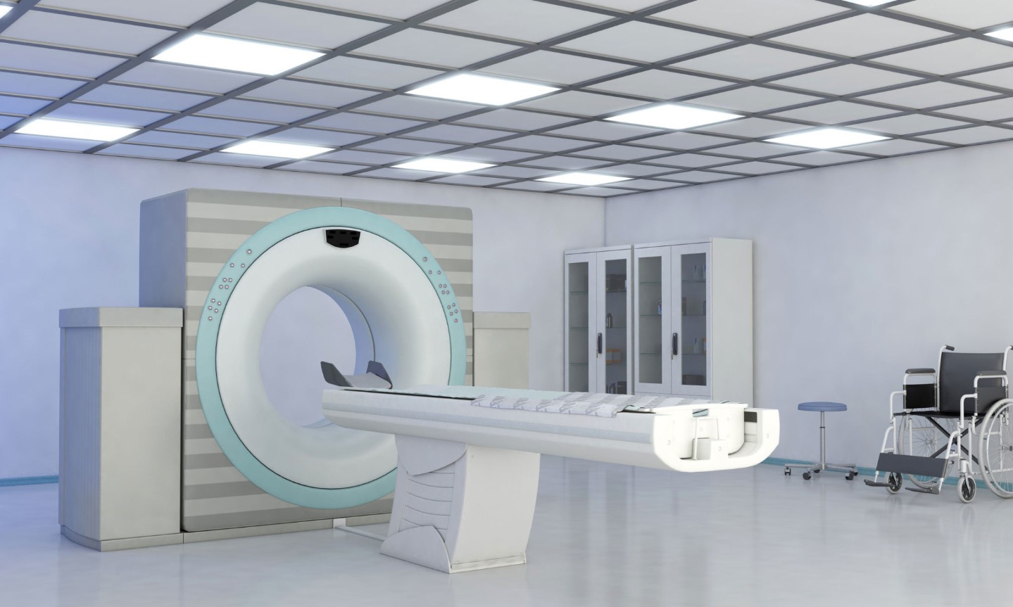
This is what went through my mind as I listened to a news story a last month. Patients receiving computed tomography (CT, or “CAT”) scans at Cedars-Sinai Medical Center in Los Angeles were exposed to several times the usual dose of radiation than is needed for the scans they received.
“Beginning in February 2008, each time a patient at Cedars-Sinai Medical Center received a CT brain perfusion scan -- a state-of-the-art procedure used to diagnose strokes -- the dose displayed would have been eight times higher than normal. No standard medical imaging procedure would use so much radiation, which one expert said is on par with the levels used to blast tumors.
“Somebody should have noticed. But nobody did -- everybody trusted the machines.
“Late last week, the U.S. Food and Drug Administration and Cedars-Sinai revealed that 206 stroke patients who received scans at the prestigious Los Angeles hospital were overdosed with radiation. Now doctors and safety experts around the country face a troubling question: In an era of supposedly fail-safe medical technology, how did the problem go undetected for 18 months?”
This story broke about the same time two studies were published in the Archives of Internal Medicine regarding the risk of cancer from radiation associated with routine CT scans. In the first paper the researchers determined that approximately 29,000 future cancers (half of which will be fatal) will arise in the United States from the radiation used in conjunction with CT scans done over the course of only one year. This is based on an assessment of the number of CT scans done during 2007 and was calculated using the theoretically established estimate of one death per 2000 scans performed. The second publication was a survey of CT scanning at four prominent California medical centers that found a remarkable 13-fold difference in the amount of radiation delivered by similar scans at these facilities. One site routinely deluged its patients with over 50 mSv of x-rays (see below to put this amount in perspective).
Some of you may be aware of these estimates, but I for one was not and found them both surprising and disturbing. I have been ordering CT scans on patients ever since I was an intern rotating through the emergency room and, while I’ve acknowledged the theoretic risk associated with the radiation from these studies, I’ve never really thought about it in such starkly objective terms.
Let’s back up a minute and review some basics about radiation dose in medical imaging. For us to understand the metrics of radiation dose let’s use as our baseline the amount of radiation you would receive from a standard chest x-ray, a simple and common study that exposes you to about 0.1 millisieverts (mSv) of ionizing radiation.
Now consider the CT scan. In order to produce its dazzling images of the inside of your body it must flood you with anywhere from 1.5 mSv (for a typical head CT) to 10 mSv (chest, abdomen and pelvis) of x-rays. That translates to the equivalent dose of radiation you would receive from undergoing a hundred chest x-rays. It has been said that the radiation exposure from a full-body CT scan (something that no radiologist I know would recommend, but a study that is nonetheless not infrequently requested by patients) is the same as standing a mile and a half away from the WWII atomic blasts in Japan.
Whether you like it or not you already receive a fairly hefty dose of unavoidable radiation on a daily basis coming at you from ordinary sources such as the sun and naturally occurring radioactive isotopes in the soil. It is estimated that each person is exposed to an average of 2.4 mSv of ionizing background radiation per year (equaling 24 chest x-rays). By undergoing a CT scan you are at least doubling your yearly dose of radioactive exposure.
Why am I telling you all this? Simply to remind you that our wonderful imaging technology is not free—it comes at a cost that is significant to at least some patients. My more frequent readers will recall a recent piece I wrote extolling the virtues of coronary CT angiography. At present this study constitutes one of the most radiation-intensive scans we can perform, zapping you with up to 13 mSv for one set of images. Based on the calculations from one of the papers I cited above, one in every 270 forty-year-old women undergoing a CT coronary angiogram will develop cancer from the procedure. This doesn’t mean we shouldn’t use this valuable tool—we just have to be very careful to apply it only to those patients who really need it.
It was December 22nd, 1895 when Wilhelm Roentgen introduced radiation-based medical imaging to the world and in the years since then this marvelous technology, for all its risk, has saved exponentially more lives than it has put in jeopardy. But for this equation to work in our favor we need to reserve the use of CT scans for only those who would truly profit from the information they provide.
The message to you as a patient is to not expect to be treated to the most technologically advanced imaging study when good old-fashioned clinical reasoning could suffice. In the end it may not be in your best interest to pester your doctor into ordering a CT scan when your clinical scenario doesn’t justify it.
And the message to us doctors is to think twice before we casually request such comprehensive radiology imaging. While killing a patient by ordering an unnecessary CT scan may not stir as much soul searching on the part of the doctor as inadvertently severing a critical artery during the course of a botched surgery, the outcome for the patient is nonetheless the same.


